What Causes Skin Cancer?
While UV radiation from the sun is the most common cause of skin cancer, there are others! Genetics, viruses (HPV, Merkel cell polyomavirus), trauma, ionizing radiation, indoor tanning, immunosuppression, certain medications, tobacco use, chronic scarring and inflammatory conditions, and chemicals (arsenic, coal tar, soot, mineral oil, nitrogen mustard, polychlorinated biphenyls, mineral oil, pesticides) may also cause skin cancers.
Types of Skin Cancer
-
Basal Cell Carcinoma
1 in 5 Americans will develop a skin cancer in their lifetime, and basal cell carcinoma is by far the most common type of skin cancer. It is slow growing and primarily locally destructive. First, the cancer destroys skin, then progresses to destroy deeper structures including bone. Basal cell carcinoma is typically not life threatening, but left untreated up to 1 in 1,000 will metastasize (spread to other parts of the body).
-
Squamous Cell Carcinoma
Squamous cell carcinoma is the second most common type of skin cancer. It may grow rapidly or slowly. Squamous cell carcinoma is typically not life threatening when treated in a timely manner. However, left untreated it has a 1-5% risk of metastasizing (spreading to other parts of the body).
-
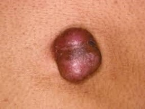
Melanoma
Melanoma is the third most common type of skin cancer. It accounts for 4% of all skin cancers. It can be more aggressive than basal cell and squamous cell carcinomas, with a higher risk of spreading to other parts of the body (metastasizing) left untreated. In addition to surgical removal, your dermatologist may recommend additional workup and treatment depending on the stage of the melanoma at diagnosis.
Less Common Skin Cancers
Prognosis and treatment depends on tumor type and characteristics. Your dermatologist and surgeon will formulate the best treatment plan for you.
Dermatofibrosarcoma Protuberans
- Atypical Fibroxanthoma
- Angiosarcoma
- Dermatofibrosarcoma Protuberans
- Extramammary Paget’s Disease
- Kaposi’s Sarcoma
- Merkel Cell Carcinoma
- Microcystic Adnexal Carcinoma
- Mucinous Carcinoma
- Other Eccrine Carcinomas
- Pleomorphic Dermal Sarcoma
- Sebaceous Carcinoma
Types of Skin Cancer Treatment
Your dermatologist and surgeon will take into account tumor type, tumor characteristics, and individual factors to select the most appropriate treatment plan for your skin cancer.
- Mohs Micrographic Surgery – Skin cancer is surgically removed in a microscopically controlled fashion and margins are checked at the same time as the surgery by the surgeon.
- Excision – Skin cancer may be cut out with a margin and sent out to a pathologist to ensure the margins are clear. The wound is typically repaired at the same time.
- Electrodessication and Curettage –Skin cancer may be “scraped” out with a sharp instrument and ”burned” by cauterization.
- Intralesional Chemotherapy – Skin cancer may be injected with chemotherapy during several sessions spaced out one to several weeks apart.
- Topical Chemotherapy – Low grade, shallow skin cancers may be treated with topical chemotherapy medications.
- Other Treatments – Other treatments for skin cancer include oral medications, injectable medications, radiation therapy, and photodynamic therapy.
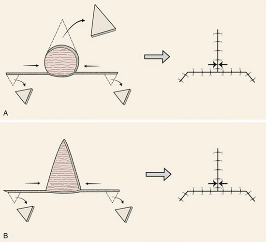
What is Mohs Micrographic Surgery?
Photo Gallery
* The following images contain graphic surgical content. Viewer discretion advised. Patient consent obtained for use of de-identified images for educational purposes.
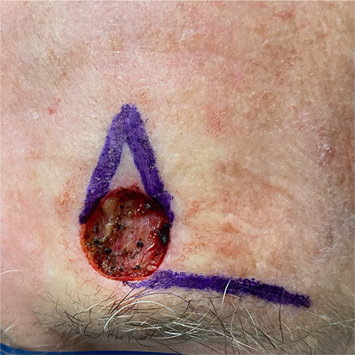
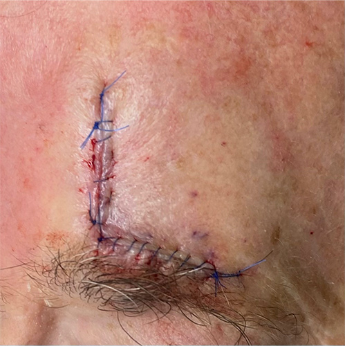
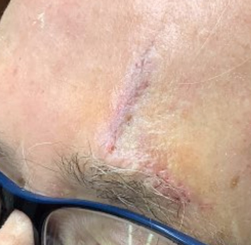
Mohs Micrographic Surgery, Skin Cancer Surgery, and Reconstructive Surgery
Face
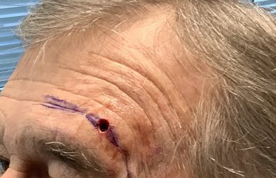
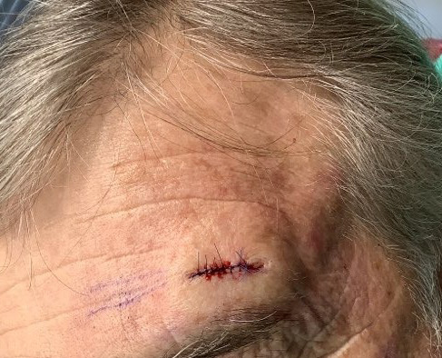
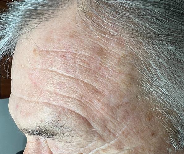
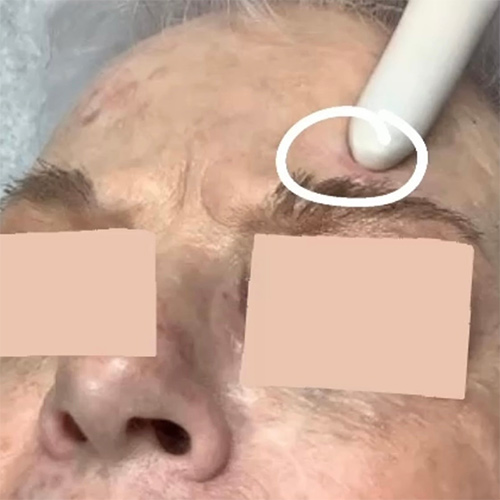
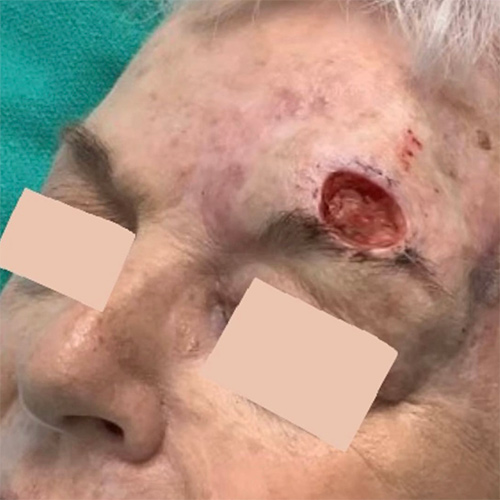
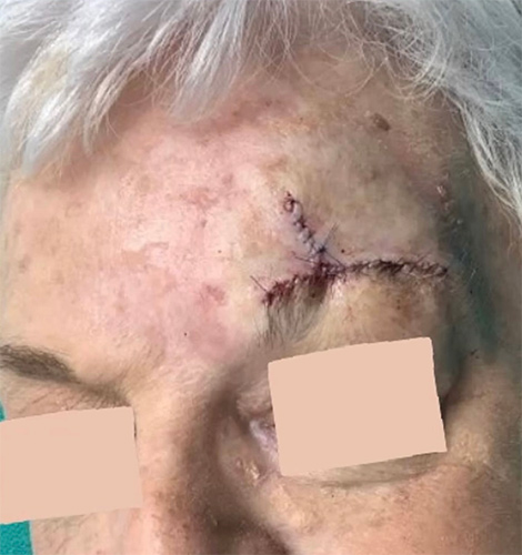
Case 1
Mohs Micrographic Surgery on the forehead + reconstructive surgery with a flap + 1 week post-op photo. The scar will become less red and puffy with time.
* Patient consent obtained for use of de-identified images for educational purposes.
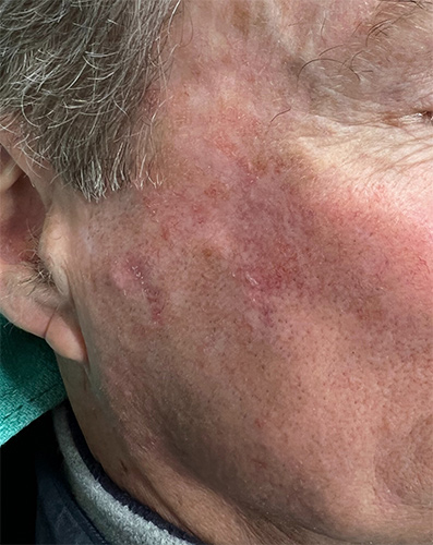
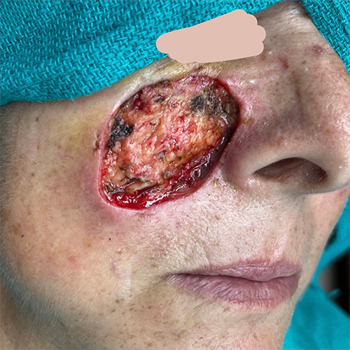
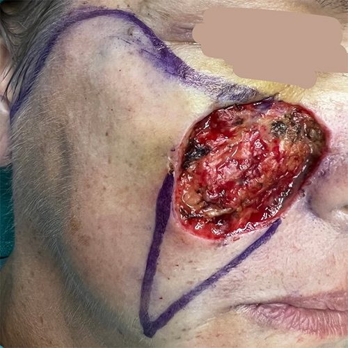
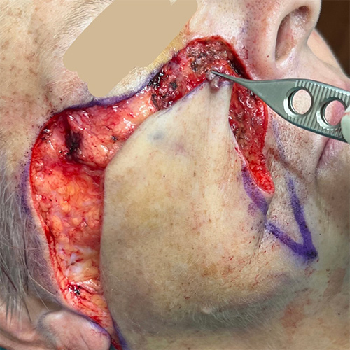
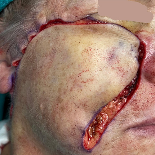
Case 2
Mohs Micrographic Surgery on the cheek + reconstructive surgery with a straight line closure + 3 month post-op photo. The scar will become even more subtle time.
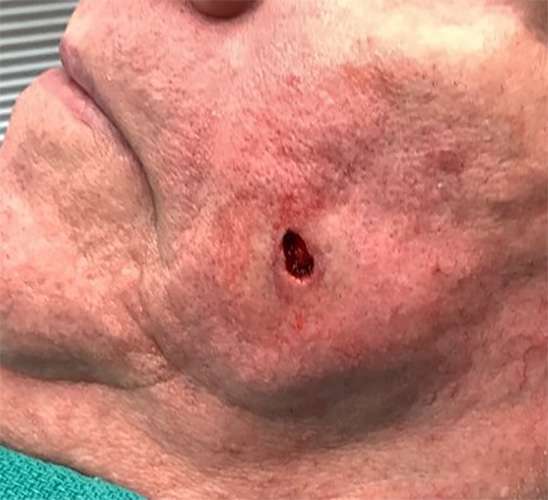
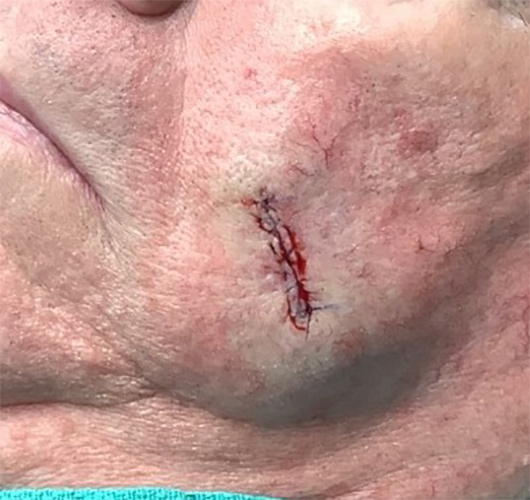
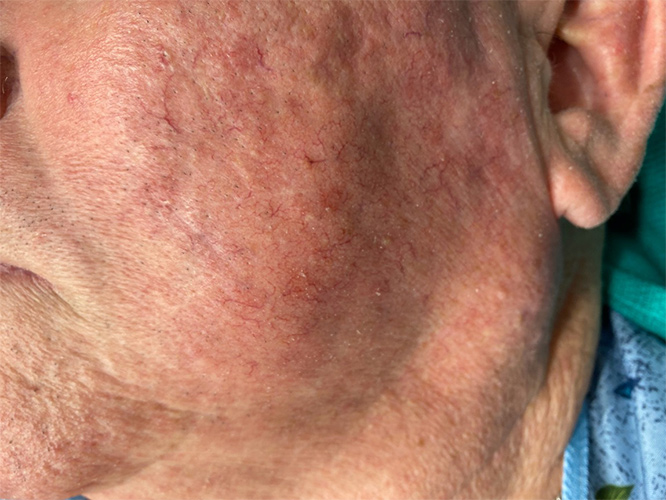
* Patient consent obtained for use of de-identified images for educational purposes.
Case 3
Mohs micrographic surgery on the cheek + reconstructive surgery with a linear closure + 1 week post-op + 12 month post-op photos
* Patient consent obtained for use of de-identified images for educational purposes.
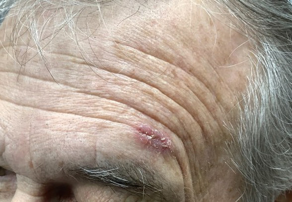
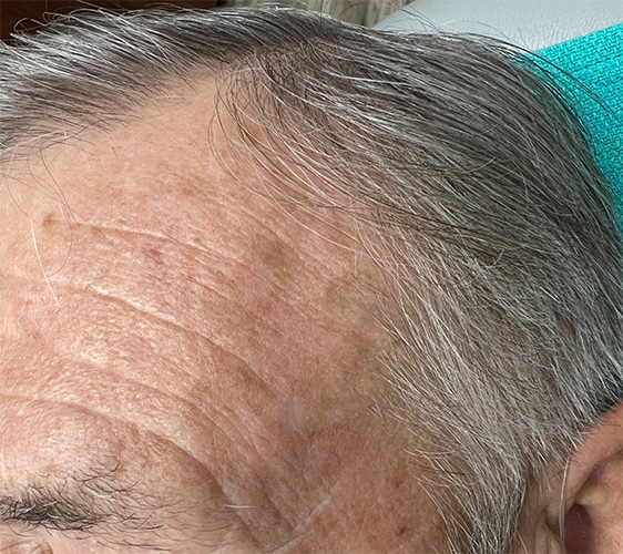
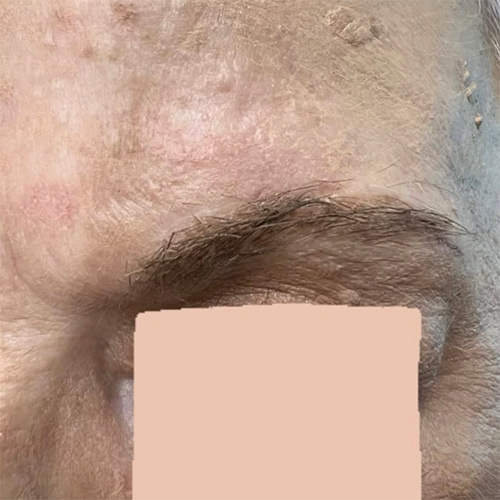
Case 4
Mohs micrographic surgery on the forehead + reconstructive surgery with a flap + 12 month post-op photos
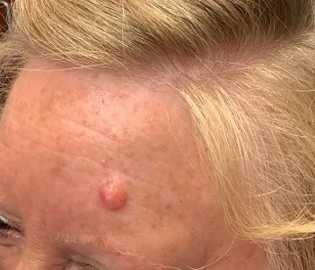
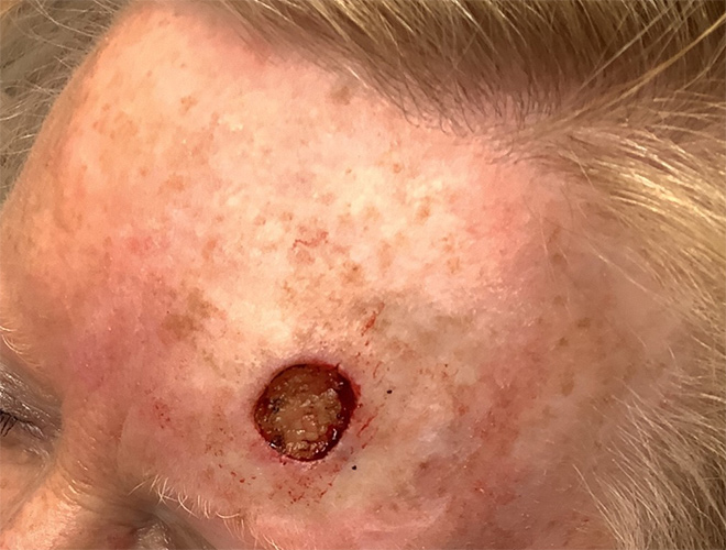
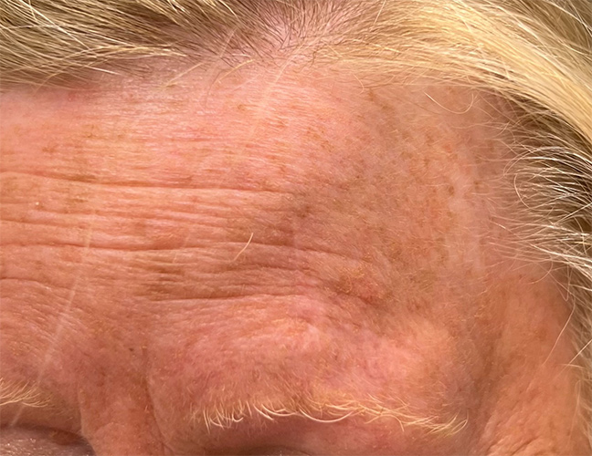
* Patient consent obtained for use of de-identified images for educational purposes.
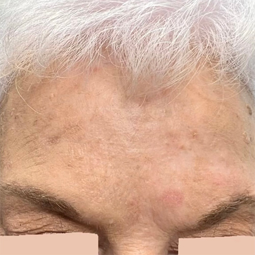
Case 5
Mohs micrographic surgery on the forehead + reconstructive surgery with a linear closure + 1 week post-op + 6 month post-op photos
* Patient consent obtained for use of de-identified images for educational purposes.
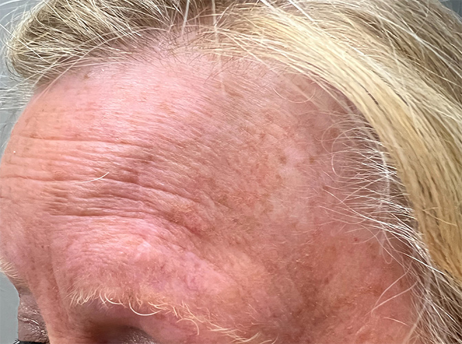
Case 6
Mohs micrographic surgery on the forehead + reconstructive surgery with a flap + 6 month post-op photos
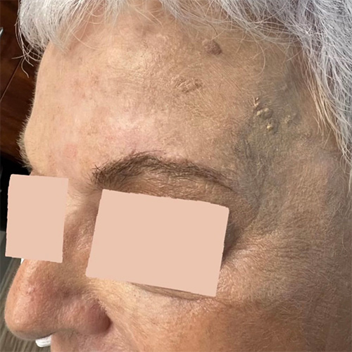
* Patient consent obtained for use of de-identified images for educational purposes.
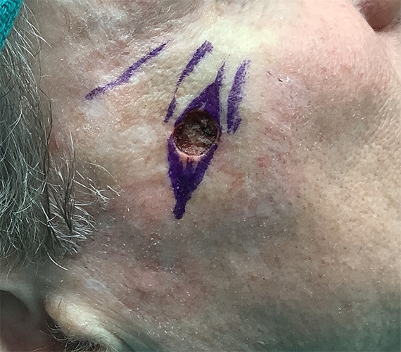
Case 7
Mohs micrographic surgery on the cheek + reconstructive surgery with a flap
* Patient consent obtained for use of de-identified images for educational purposes.
Case 8
Mohs micrographic surgery on the nasal crease + reconstructive surgery with a linear closure + 12 month post-op photo
* Patient consent obtained for use of de-identified images for educational purposes.
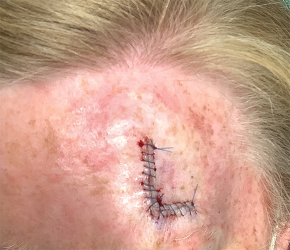
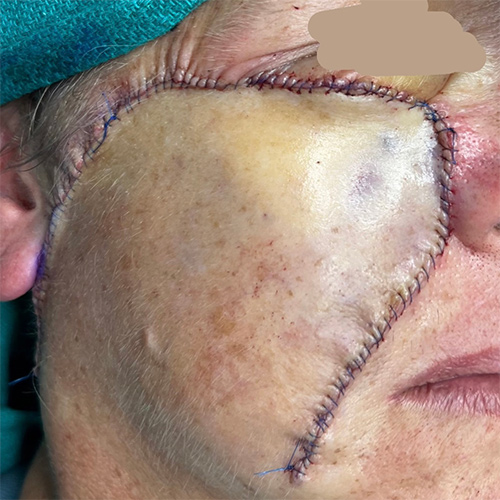
Scalp
Case 1
Skin cancer excision + reconstructive surgery with a flap + 2 week post-op photo. The scar will become less red and puffy with time.
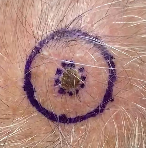
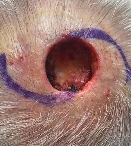
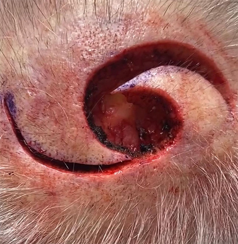
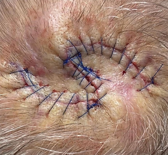
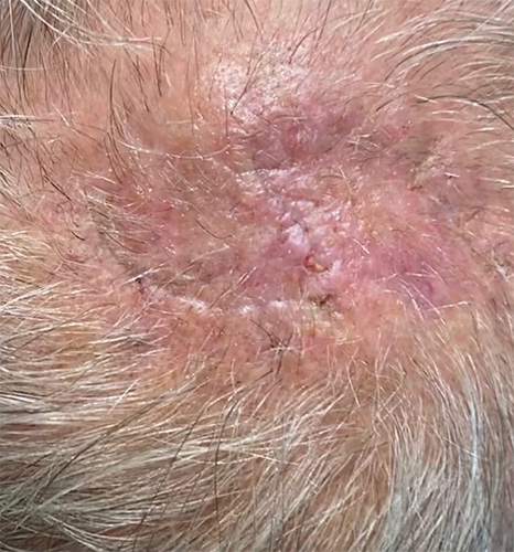
* Patient consent obtained for use of de-identified images for educational purposes.
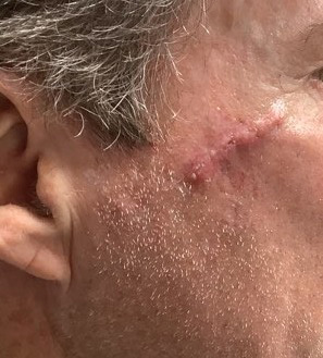
Neck
Case 1
Mohs Micrographic Surgery on the ear and neck + reconstructive surgery with straight line closures + 10 month post-op photos.
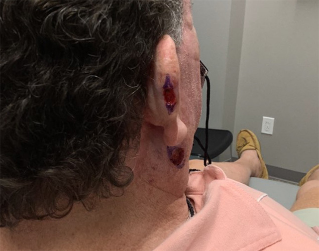
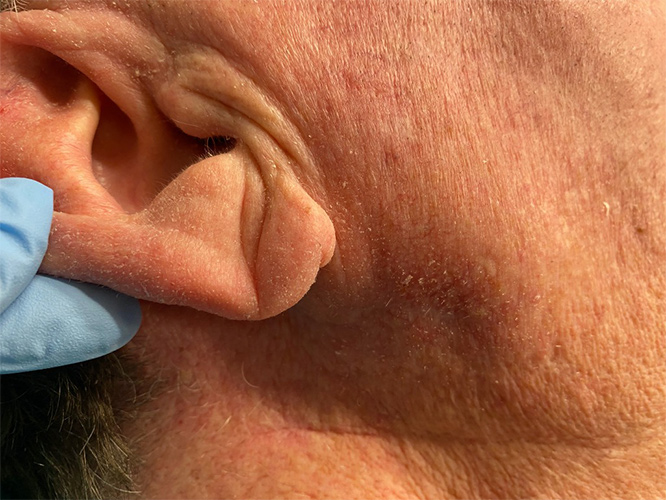
* Patient consent obtained for use of de-identified images for educational purposes.
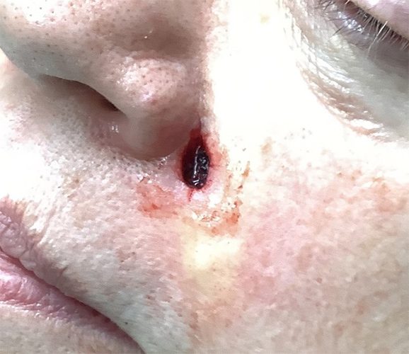
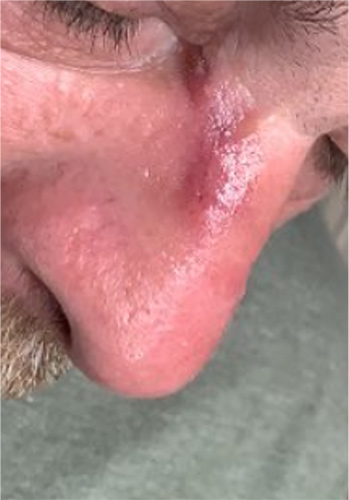
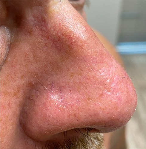
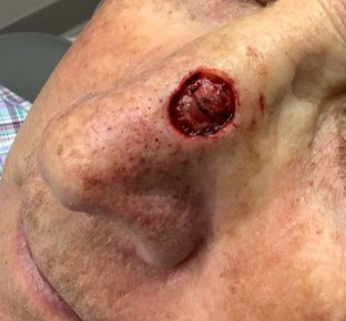
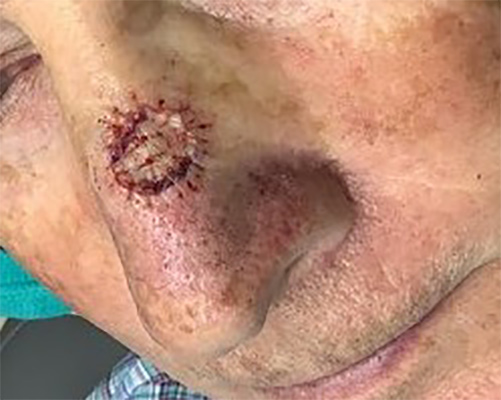
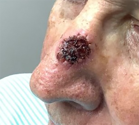
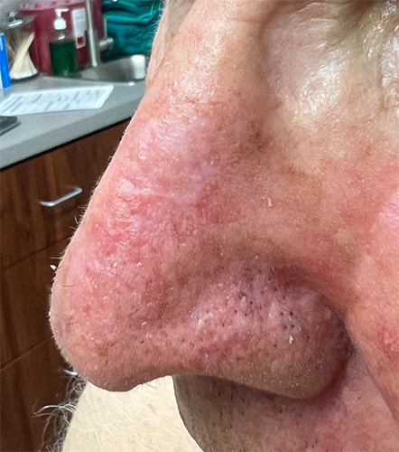
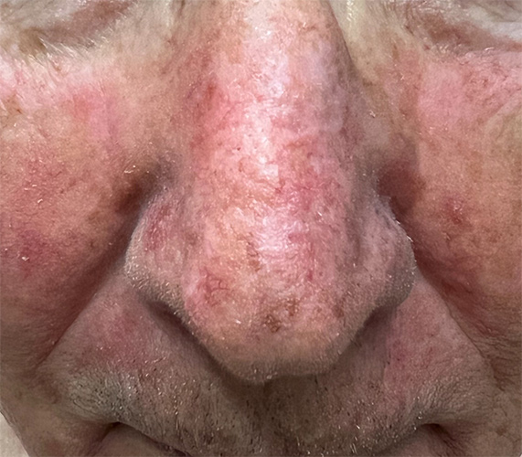
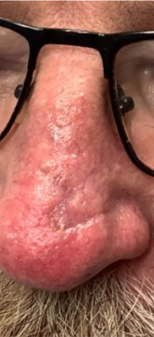
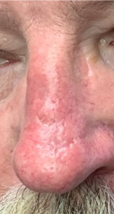
Nose
Case 1
Mohs Micrographic Surgery on the Nose + reconstructive surgery with a flap + 1 week post-op + 4 month post-op photos
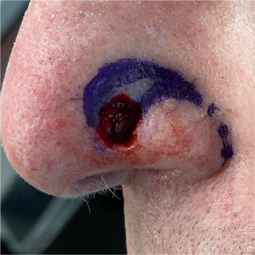
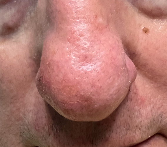
* Patient consent obtained for use of de-identified images for educational purposes.
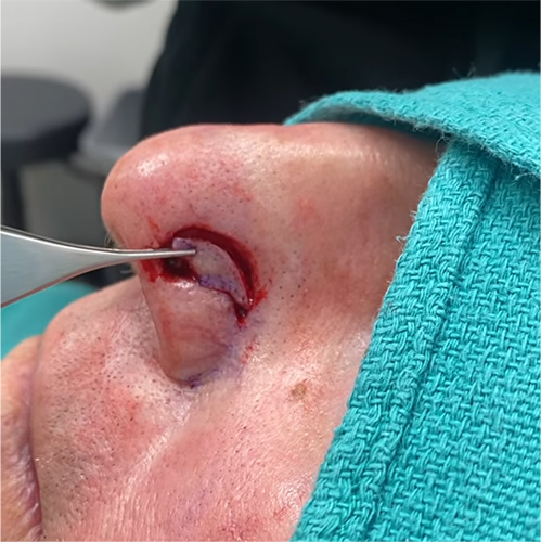
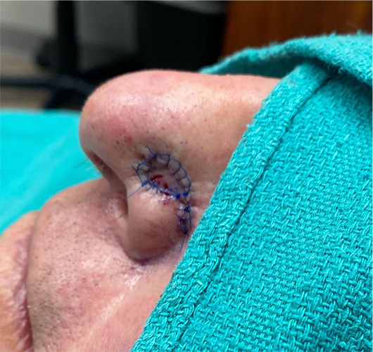
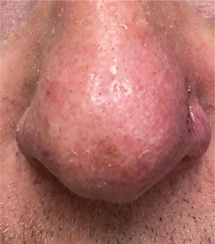
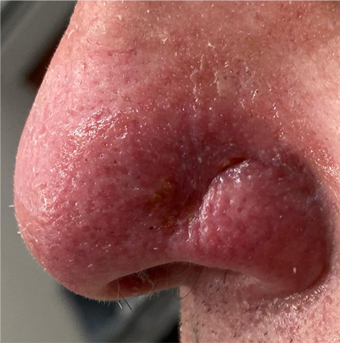
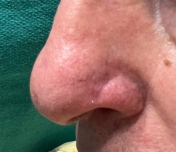
Case 2
Mohs Micrographic Surgery on the nose + reconstructive surgery with a straight line closure + 1 week post-op photo + 2 month post-op photo.
* Patient consent obtained for use of de-identified images for educational purposes.
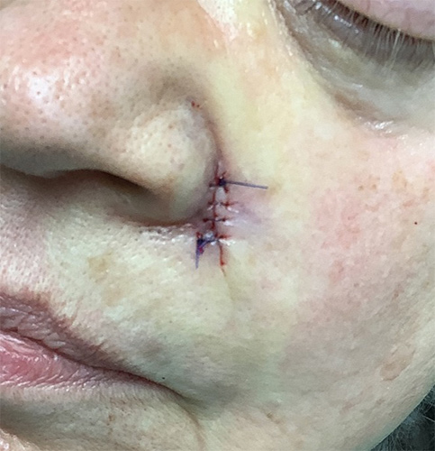
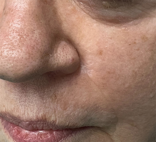
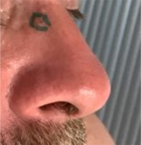
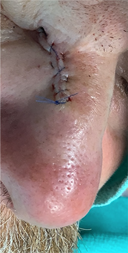
Case 3
Mohs micrographic surgery on the nose + reconstructive surgery with a full thickness skin graft + 1 week post-op + 12 month post-op photos
* Patient consent obtained for use of de-identified images for educational purposes.
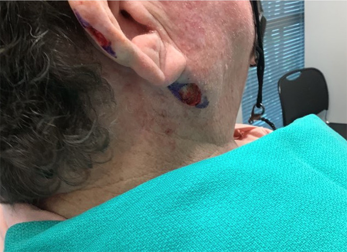
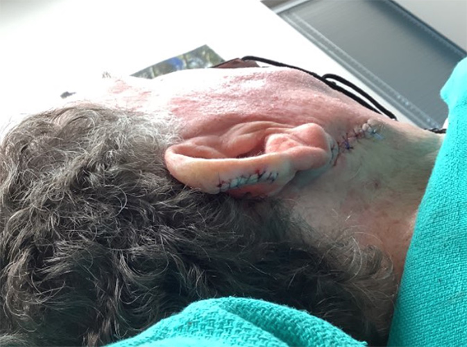
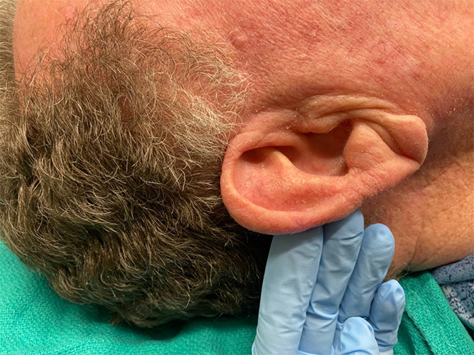
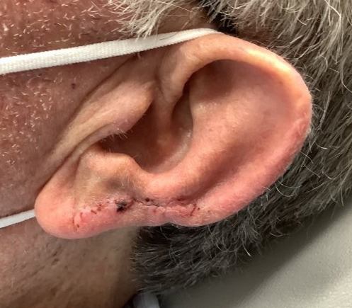
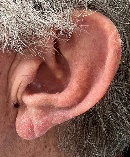
Ears
Case 1
Mohs Micrographic Surgery on the ear + reconstructive surgery with a flap + 2 week post-op photos. The scar will become less red and puffy with time.
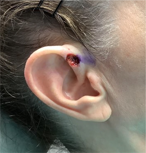
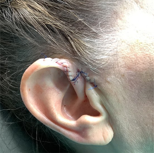
* Patient consent obtained for use of de-identified images for educational purposes.
Case 2
Mohs Micrographic Surgery on the ear and neck + reconstructive surgery with straight line closures + 10 month post-op photos.
* Patient consent obtained for use of de-identified images for educational purposes.
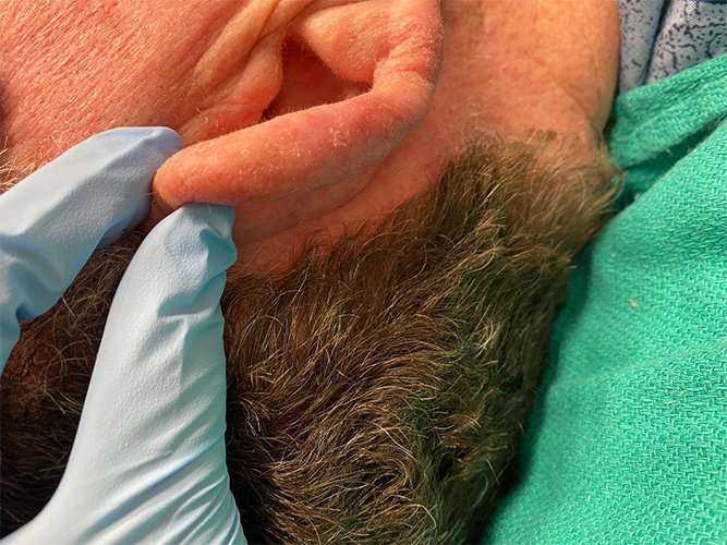
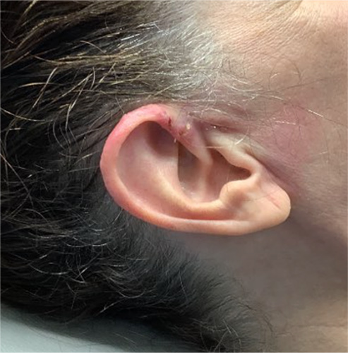
Case 3
Mohs micrographic surgery on the ear + reconstructive surgery with a linear closure + 1 week post-op + 9 month post-op photos
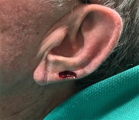
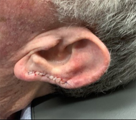
* Patient consent obtained for use of de-identified images for educational purposes.
Scar Revision
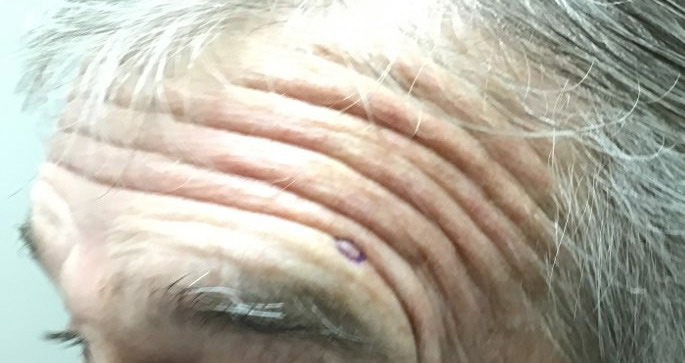
Scar Revision - Nose
Case 1
Depressed scar greatly improved after treatment with just one session of microneedling.
*Patient consent obtained for use of de-identified images for educational purposes.