Reconstructive Surgery Following Mohs Micrographic Surgery
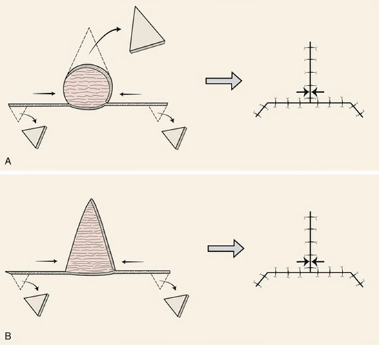
After skin cancer is treated surgically, the wound is most often repaired on the same day by your skin cancer surgeon. Wounds are closed under local anesthesia. The wound may be closed in a straight line (also known as primary closure), with a flap (transfer of adjacent skin), or with a graft (transfer of distant skin and sometimes structural cartilage). There are innumerable subtypes of these repairs. In some cases the wound is left to heal on its own (sometimes with the help of special wound dressings or skin substitutes) in a process called second intention healing. Your surgeon will choose the repair that maintains function, optimizes appearance, and satisfies patient preferences.
Photo Gallery
* The following images contain graphic surgical content. Viewer discretion advised. Patient consent obtained for use of de-identified images for educational purposes.
Mohs Micrographic Surgery, Skin Cancer Surgery, and Reconstructive Surgery
Face
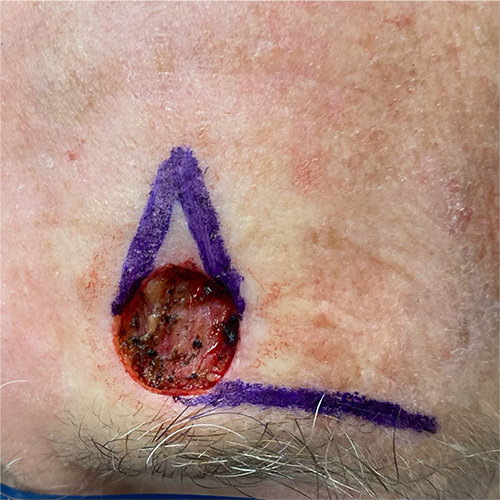
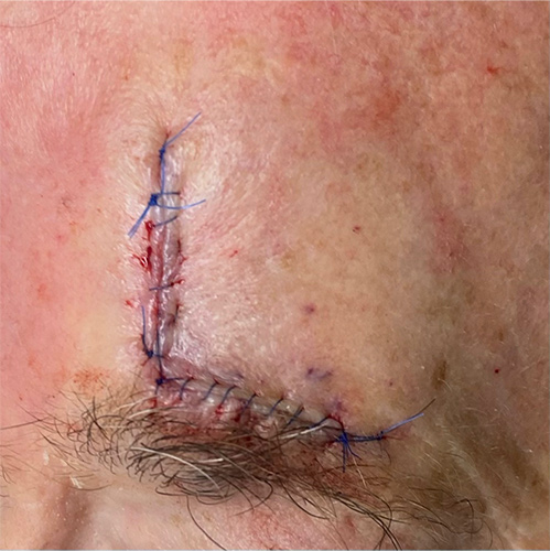
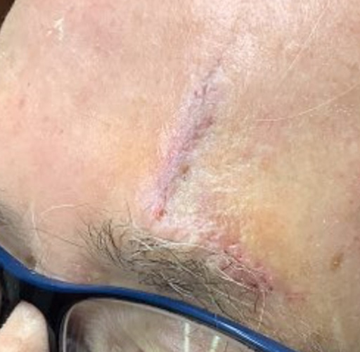
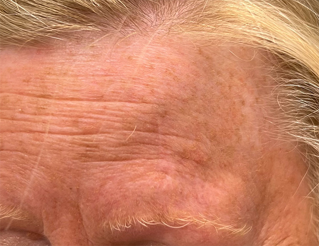
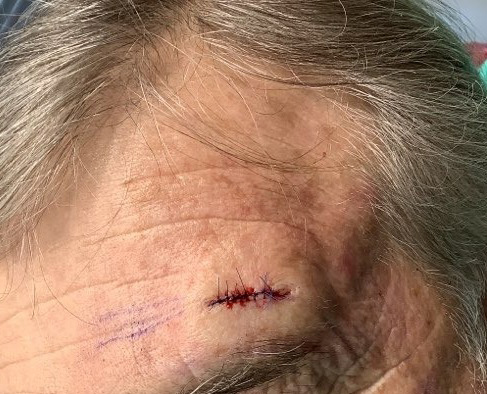
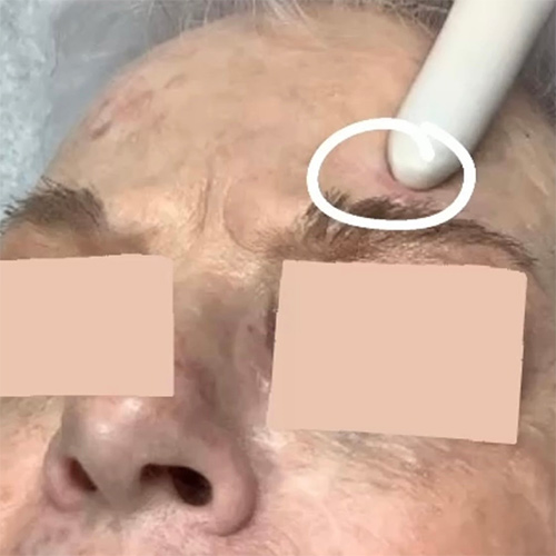
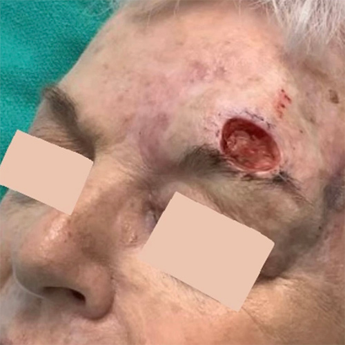
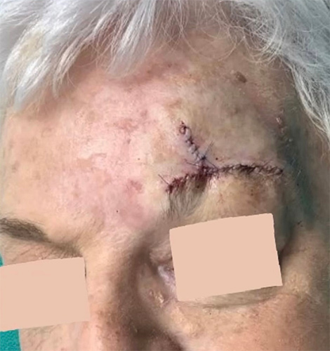
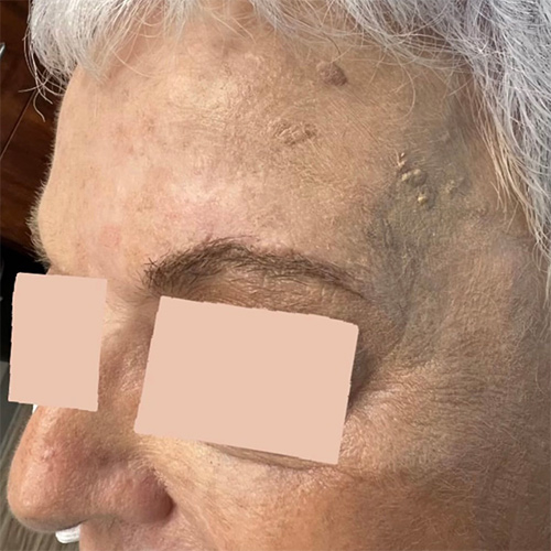
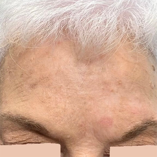
Case 1
Mohs Micrographic Surgery on the forehead + reconstructive surgery with a flap + 1 week post-op photo. The scar will become less red and puffy with time.
* Patient consent obtained for use of de-identified images for educational purposes.
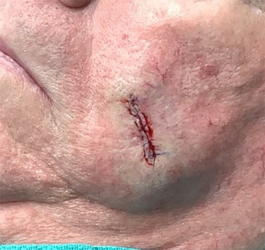
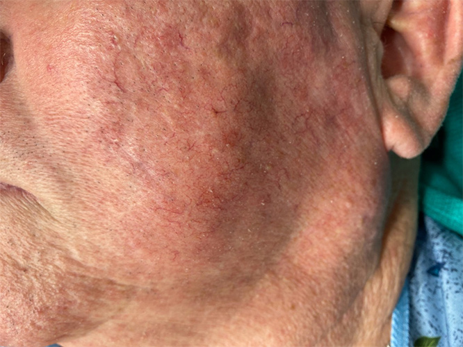
Case 2
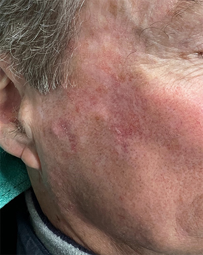
Mohs Micrographic Surgery on the cheek + reconstructive surgery with a straight line closure + 3 month post-op photo. The scar will become even more subtle time.
* Patient consent obtained for use of de-identified images for educational purposes.
Case 3
Mohs micrographic surgery on the cheek + reconstructive surgery with a linear closure + 1 week post-op + 12 month post-op photos
* Patient consent obtained for use of de-identified images for educational purposes.
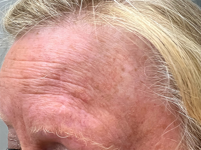
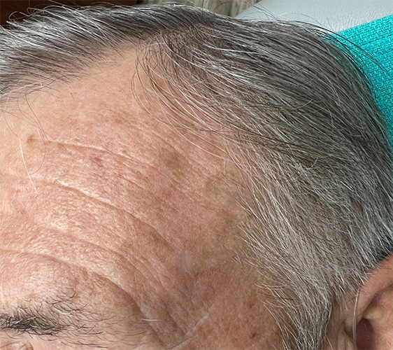
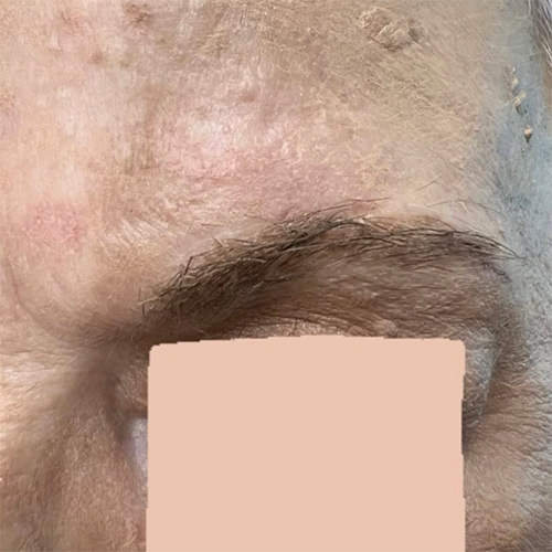
Case 4
Mohs micrographic surgery on the forehead + reconstructive surgery with a flap + 12 month post-op photos
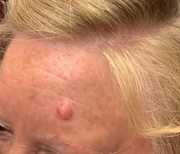
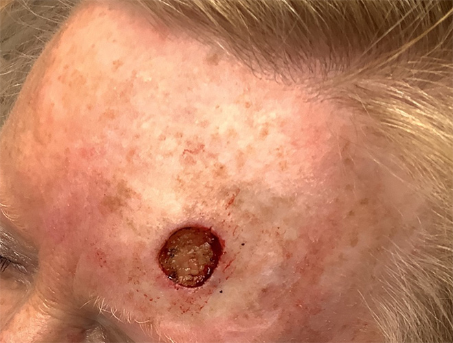
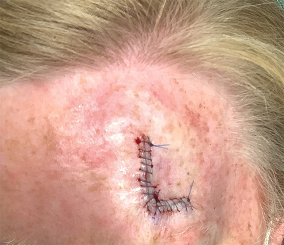
* Patient consent obtained for use of de-identified images for educational purposes.
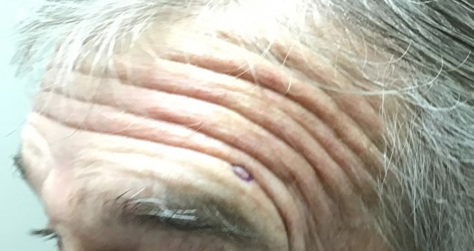
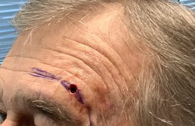
Case 5
Mohs micrographic surgery on the forehead + reconstructive surgery with a linear closure + 1 week post-op + 6 month post-op photos
* Patient consent obtained for use of de-identified images for educational purposes.
Case 6
Mohs micrographic surgery on the forehead + reconstructive surgery with a flap + 6 month post-op photos
* Patient consent obtained for use of de-identified images for educational purposes.
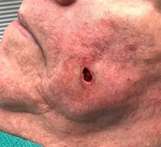
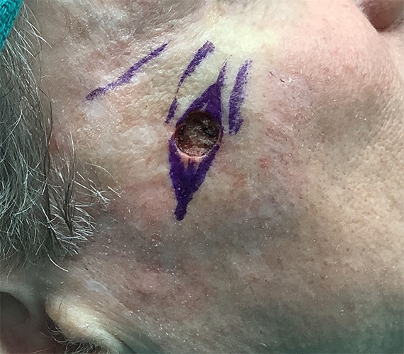
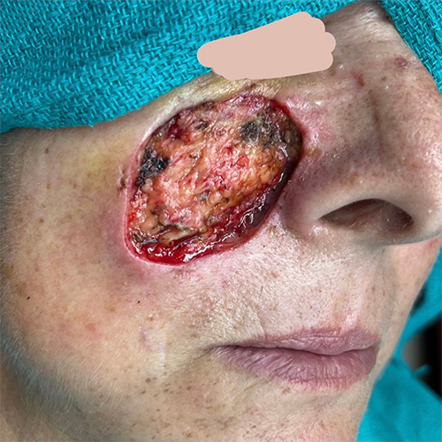
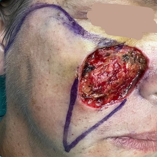
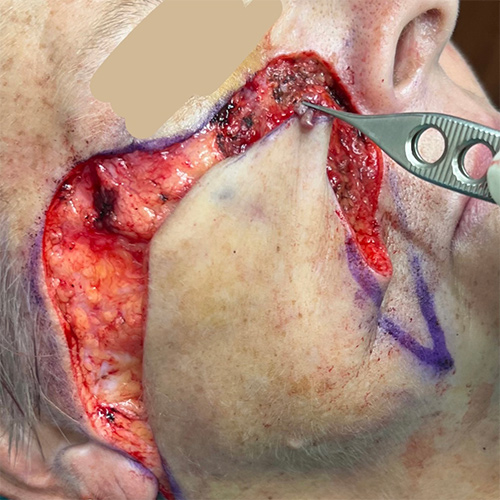
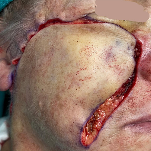
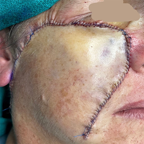
Case 7
Mohs micrographic surgery on the cheek + reconstructive surgery with a flap
* Patient consent obtained for use of de-identified images for educational purposes.
Case 8
Mohs micrographic surgery on the nasal crease + reconstructive surgery with a linear closure + 12 month post-op photo
* Patient consent obtained for use of de-identified images for educational purposes.
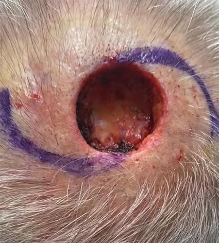
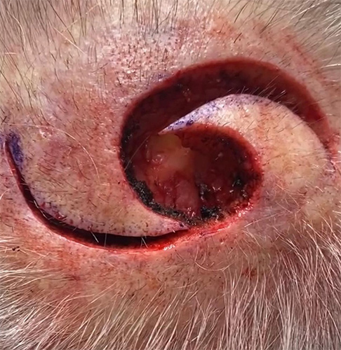
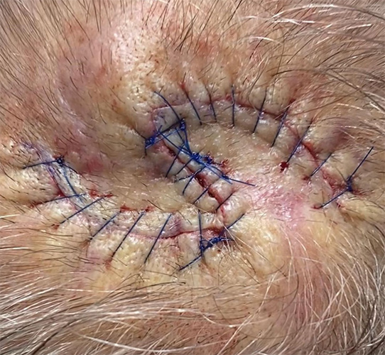
Scalp
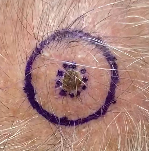
Case 1
Skin cancer excision + reconstructive surgery with a flap + 2 week post-op photo. The scar will become less red and puffy with time.
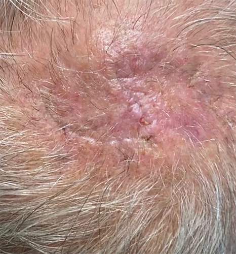
* Patient consent obtained for use of de-identified images for educational purposes.
Neck
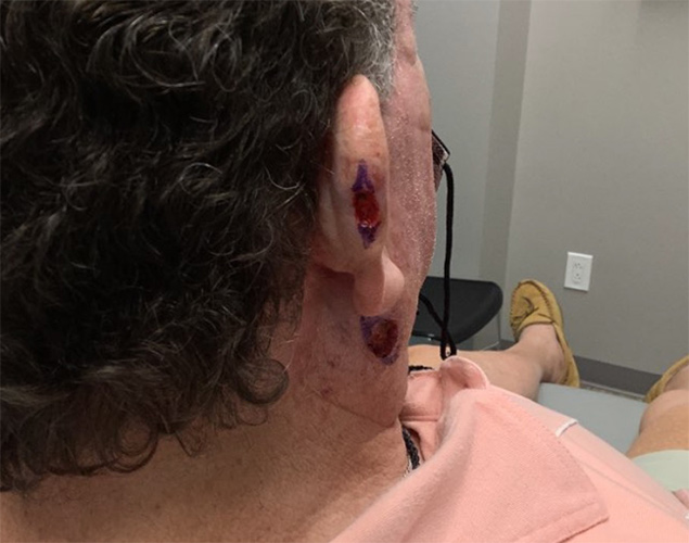
Case 1
Mohs Micrographic Surgery on the ear and neck + reconstructive surgery with straight line closures + 10 month post-op photos.
* Patient consent obtained for use of de-identified images for educational purposes.
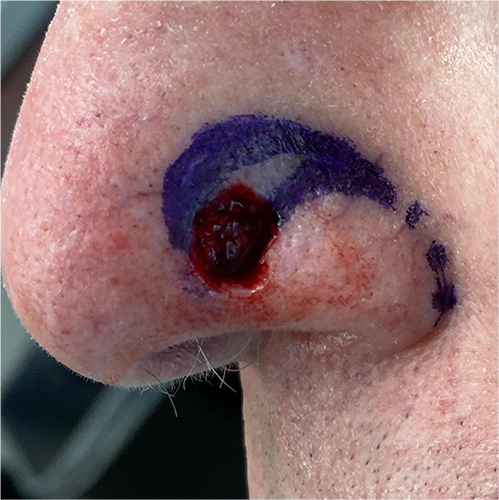
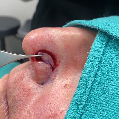
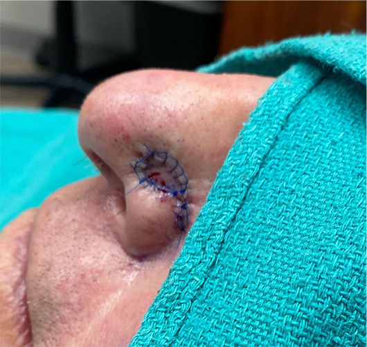
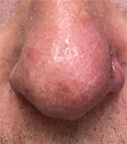
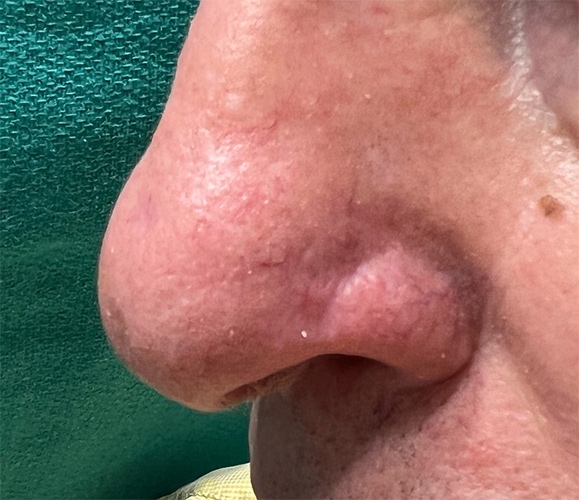
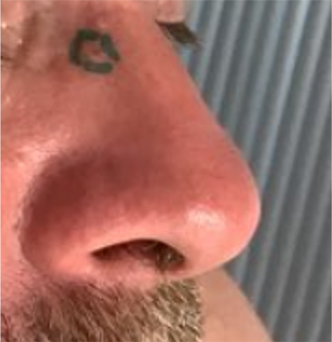
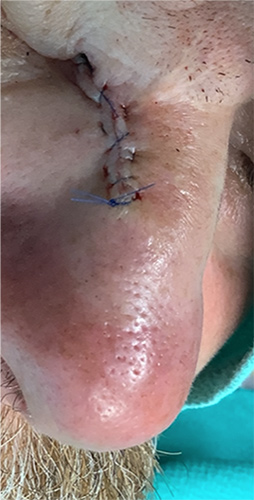
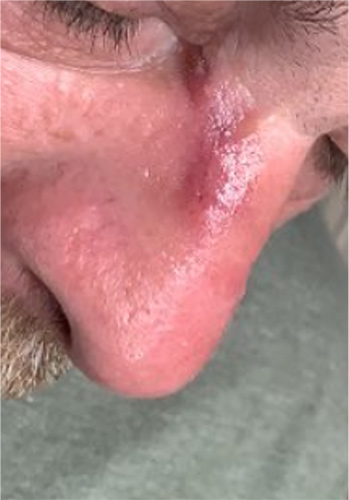
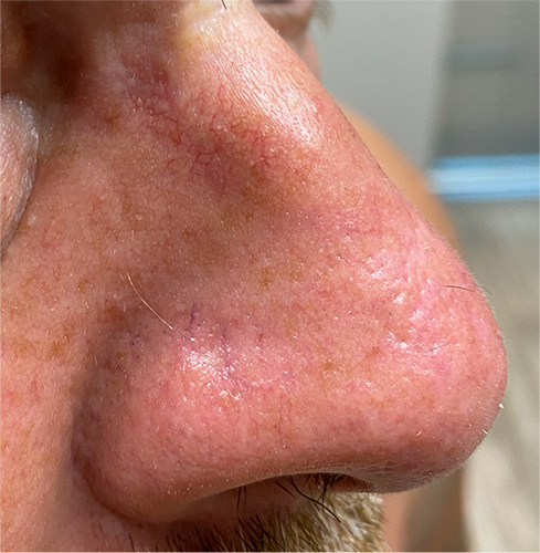
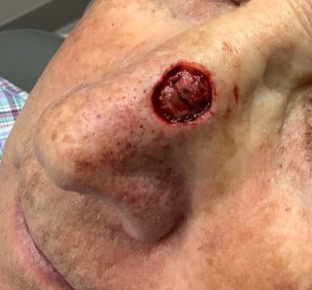
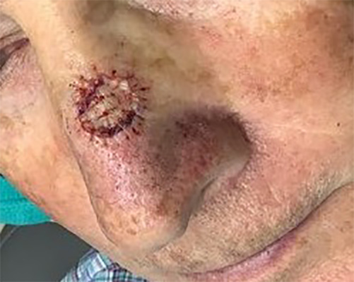
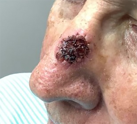
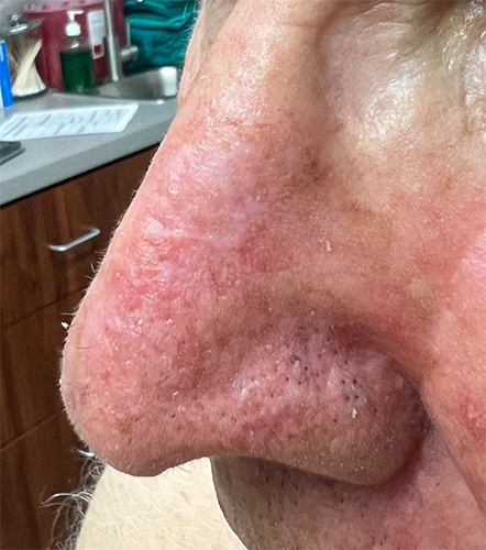
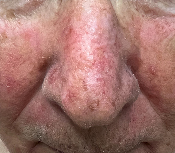
Nose
Case 1
Mohs Micrographic Surgery on the Nose + reconstructive surgery with a flap + 1 week post-op + 4 month post-op photos
* Patient consent obtained for use of de-identified images for educational purposes.
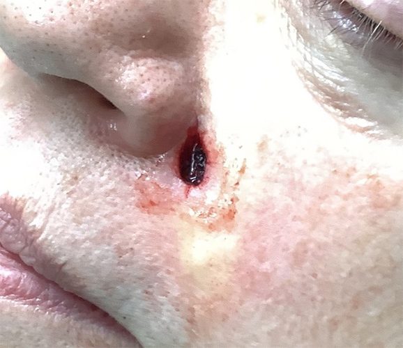
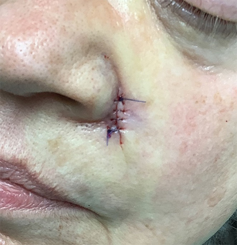
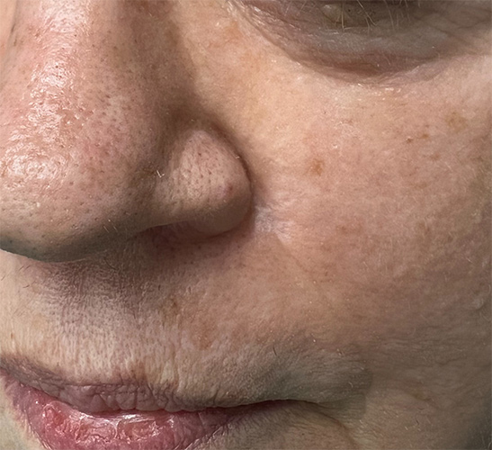
Case 2
Mohs Micrographic Surgery on the nose + reconstructive surgery with a straight line closure + 1 week post-op photo + 2 month post-op photo.
* Patient consent obtained for use of de-identified images for educational purposes.
Case 3
Mohs micrographic surgery on the nose + reconstructive surgery with a full thickness skin graft + 1 week post-op + 12 month post-op photos
* Patient consent obtained for use of de-identified images for educational purposes.
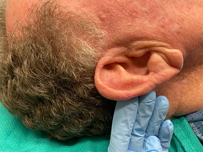
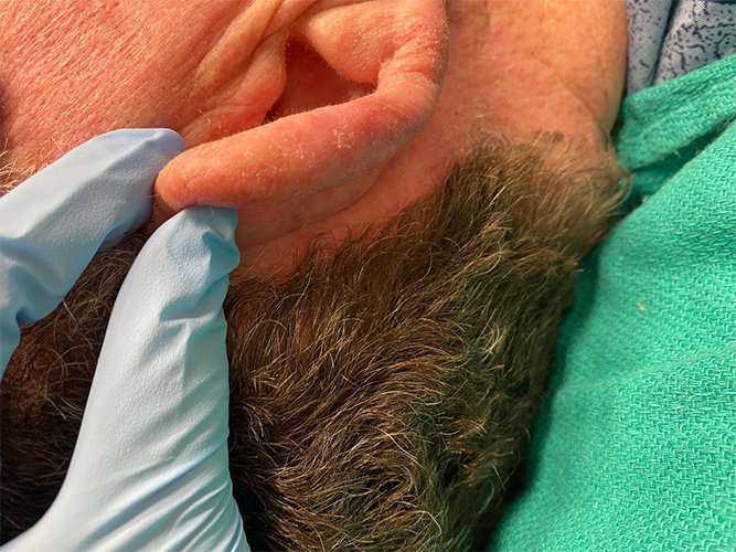
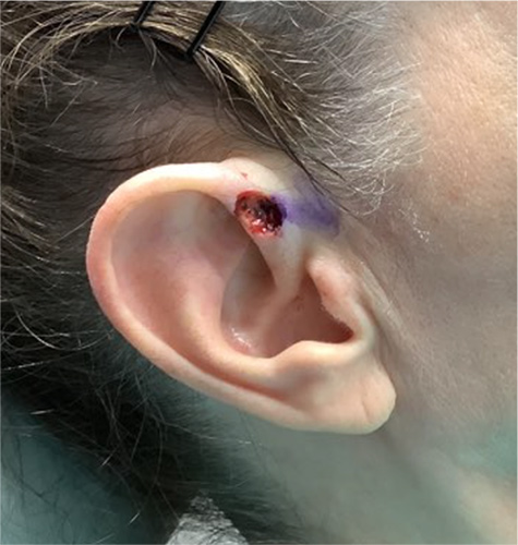
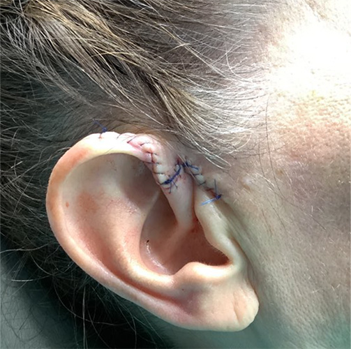
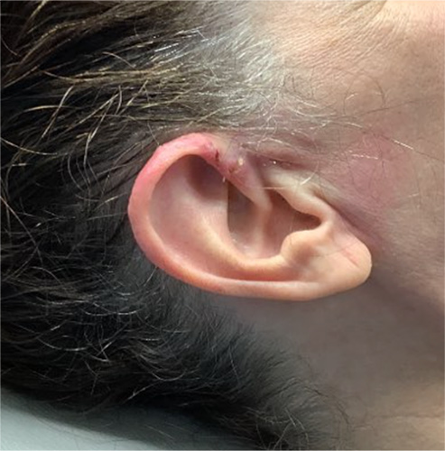
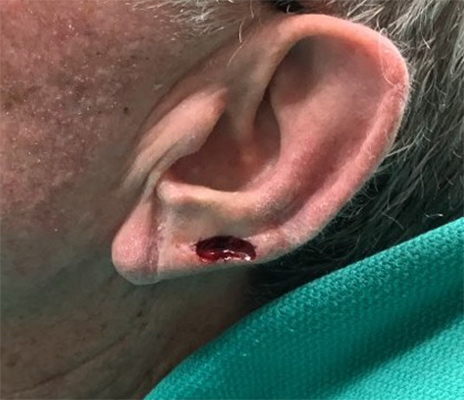
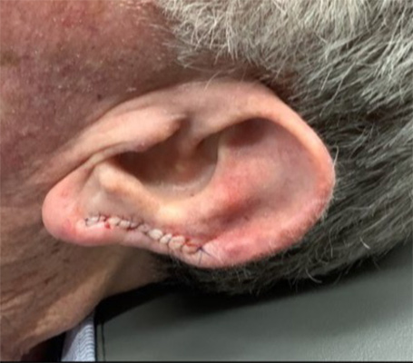
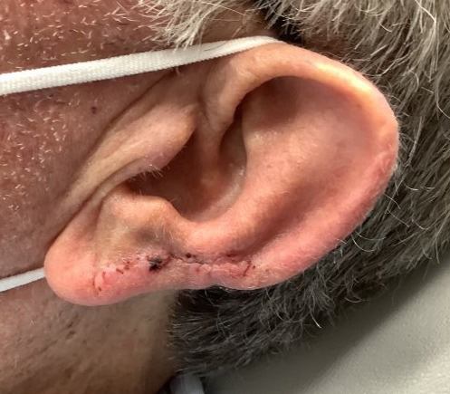
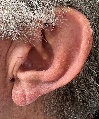
Ears
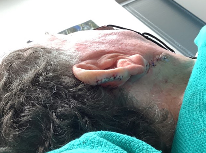
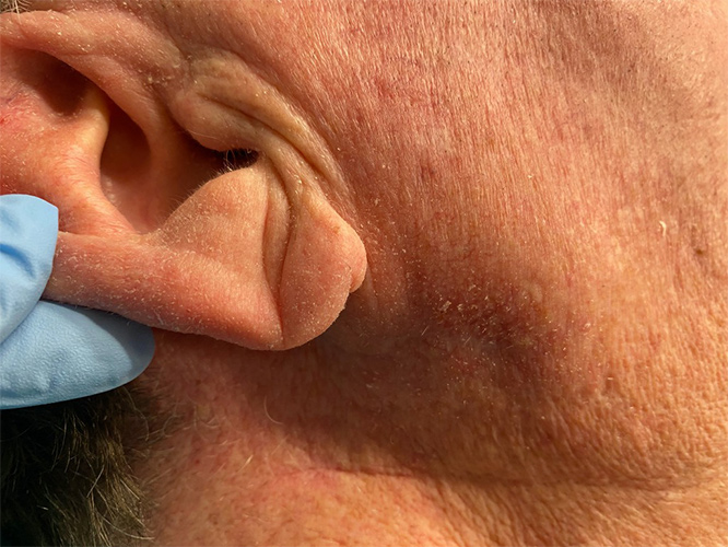
Case 1
Mohs Micrographic Surgery on the ear + reconstructive surgery with a flap + 2 week post-op photos. The scar will become less red and puffy with time.
* Patient consent obtained for use of de-identified images for educational purposes.
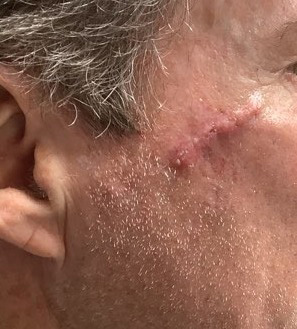
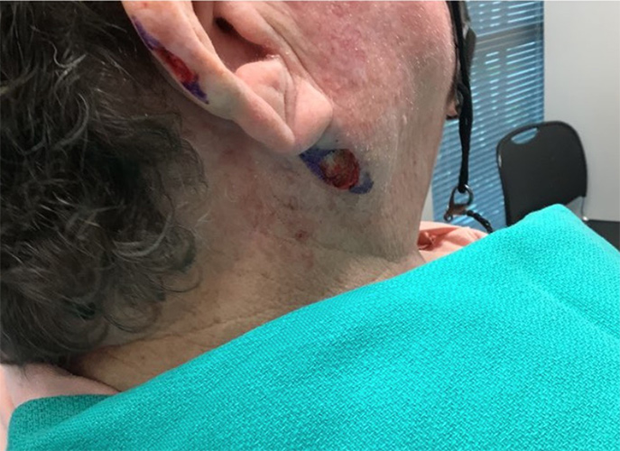
Case 2
Mohs Micrographic Surgery on the ear and neck + reconstructive surgery with straight line closures + 10 month post-op photos.
* Patient consent obtained for use of de-identified images for educational purposes.
Case 3
Mohs micrographic surgery on the ear + reconstructive surgery with a linear closure + 1 week post-op + 9 month post-op photos
* Patient consent obtained for use of de-identified images for educational purposes.
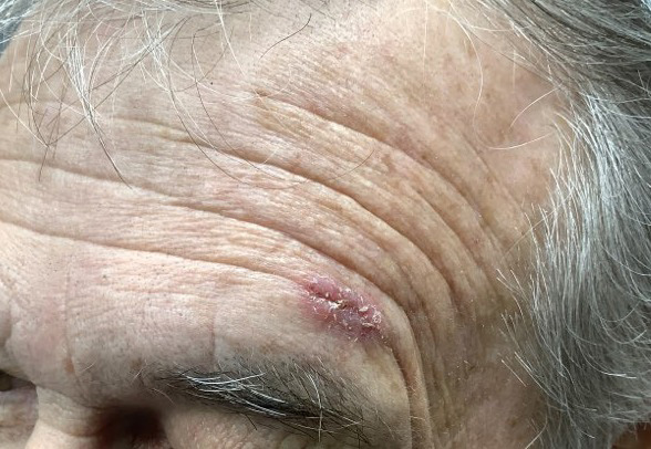
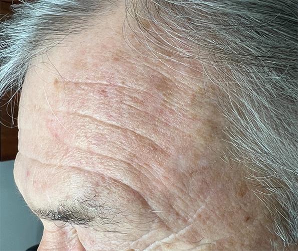
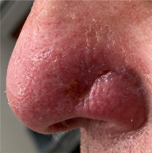
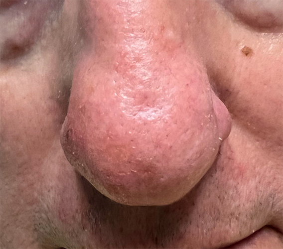
Scar Revision
Scar Revision - Nose
Case 1
Depressed scar greatly improved after treatment with just one session of microneedling.
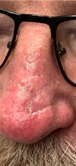
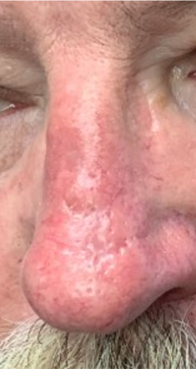
*Patient consent obtained for use of de-identified images for educational purposes.